


Specialized Diagnostic Imaging
Leaders in Advanced Veterinary Dental Diagnostic Imaging
Effective dental care relies on evaluating structures that cannot be seen during a standard oral examination. In fact, up to 70% of a tooth’s structure lies beneath the gumline, making advanced imaging an essential component of accurate diagnosis and treatment planning.
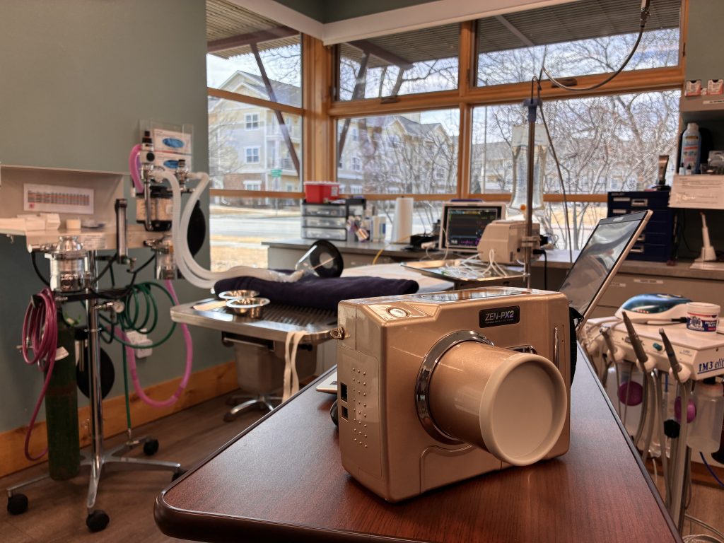
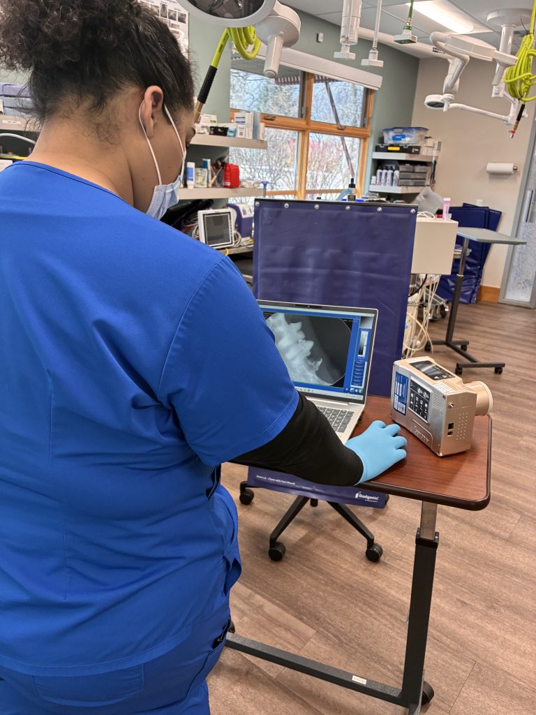
For many years, full-mouth dental radiographs have been considered the standard of care in veterinary dentistry. Dental radiographs provide valuable insight into conditions such as periodontal disease, tooth root infections, root fractures, and other abnormalities that are not visible during a routine oral examination. However, traditional dental radiographs provide only a two-dimensional representation of complex oral structures.
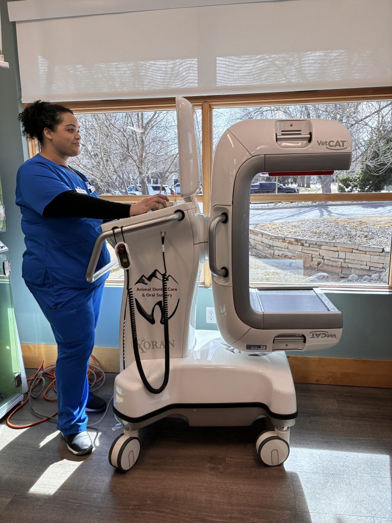
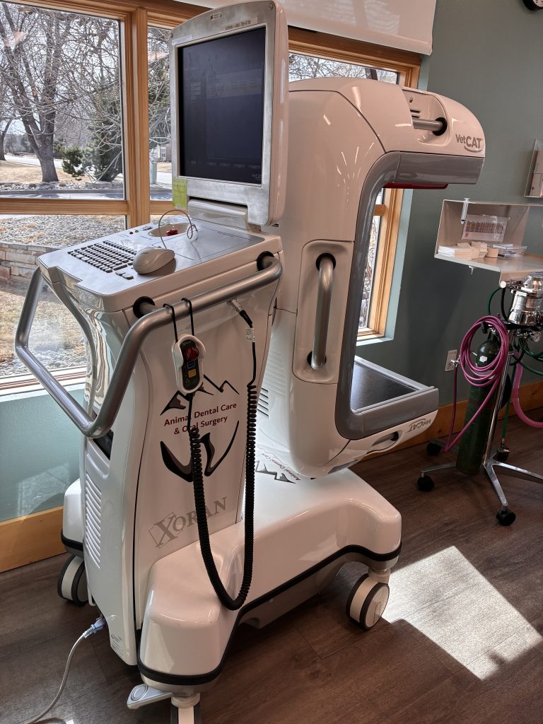
At Animal Dental Care & Oral Surgery, we are committed to utilizing the most advanced diagnostic tools available. As one of the first veterinary practices in Colorado to adopt Cone Beam Computed Tomography (CBCT) technology, we can provide an exceptional level of diagnostic accuracy and treatment planning for our patients.
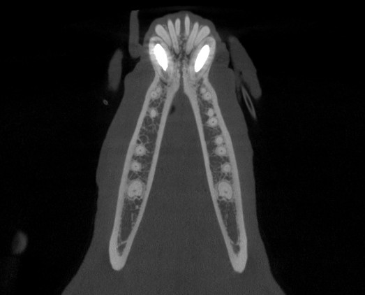
CBCT technology provides highly detailed 3D images of the teeth, jaws, skull, and surrounding structures, allowing for more accurate diagnosis than traditional dental radiographs. Our advanced Cone Beam CT unit completes a scan in approximately 30 seconds, reducing anesthesia time while helping identify periodontal disease, root resorption, endodontic disease, oral tumors, TMJ disorders, and other complex conditions. It also provides critical information for precise treatment and surgical planning.
By combining advanced imaging technology with specialized expertise in veterinary dentistry and oral surgery, we deliver more accurate diagnoses, personalized treatment plans, and exceptional patient care.