
15 Mar Variations in Tooth Anatomy in Dogs
Not every dog and cat have teeth that are just like a picture in a textbook. Some animals are born with extra teeth, missing teeth, malformed teeth, extra roots and/or teeth growing in the wrong place. Sometimes these abnormal teeth cause problems, but sometimes they are just incidental findings that illustrate how unique each pet is. Our post will discuss the variations in tooth anatomy in dogs.
Adult Dog Teeth
Dogs have a great variety of shapes in their 42 teeth. They have teeth with single roots (canines, incisors, first premolars and third molars), double roots (upper second and third premolars, lower premolars and first and second molars) and triple roots (upper fourth premolars and molars). Cats have only 30 teeth and the only triple rooted teeth are the upper fourth premolars. All the rest are single or double rooted.
Cats rarely have abnormalities in tooth shape and number, but sometimes dogs can be a little more creative. It is not unusual to find a triple rooted upper third premolar in dogs like this one. These supernumerary roots are usually bilateral, meaning if present on one side, the same tooth on the other side is likely to have it as well.
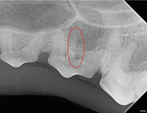
Extra roots are generally not a problem unless the tooth needs to be extracted. In that case, the veterinarian needs to be sure to get all three roots.
Puppy Teeth
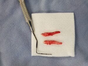
The puppy in the photo below came to visit us because he had extra teeth growing on the inside of his lower canines. Extra teeth are called “supernumerary” teeth. And although it is obvious that something is different about this puppy, we did not know if those extra teeth were a second set of canine teeth or incisors (the smaller teeth between the canines).
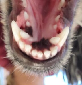
Fortunately, a dental radiograph could easily tell the difference. The extra teeth were supernumerary third incisors. Some dogs have room for extra teeth. If the extra teeth don’t cause any crowding and don’t interfere with the mouth closing, they can stay in place. In this puppy’s situation, the extra teeth were very crowded with the canines. Crowded teeth are predisposed to periodontal disease, so the extra incisors were extracted. With regular home and professional dental care, Cooper’s remaining teeth should stay healthy.
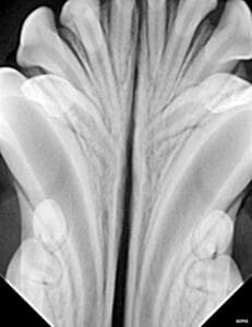
Even though incisors look pretty small, their roots are quite long. These teeth had to be extracted very carefully to avoid damage to all the other teeth nearby.
Diagnostic Imaging in Fort Collins
There’s something fascinating about variations in normal tooth anatomy. These cases illustrate the importance of diagnostic imaging such as dental radiographs and cone beam CT. At Animal Dental Care & Oral Surgery in Fort Collins, every tooth in every patient is carefully examined to determine if it is healthy and to document any abnormalities. If you think your pet has a “special” tooth, give us a call to schedule an exam!
Photo by JacLou DL from Pexels